
What Is Cataract Surgery?
Cataract surgery is the most commonly performed surgical procedure in the United States, with more than 4 million operations performed annually. The procedure involves removing the clouded natural lens of the eye and replacing it with a clear artificial intraocular lens (IOL).
The modern technique, called phacoemulsification (or "phaco"), uses ultrasound energy to break up the clouded lens into tiny fragments, which are then gently suctioned out. The entire procedure typically takes 15 to 20 minutes per eye and is performed under local anesthesia on an outpatient basis.
Before Surgery: The Pre-Operative Process
Comprehensive Eye Examination
Your ophthalmologist will perform a thorough pre-operative evaluation that includes:
- Biometry — Precise measurements of your eye's axial length and corneal curvature to calculate the correct IOL power
- Corneal topography — Detailed mapping of your corneal surface
- Endothelial cell count — Assessment of the cornea's inner cell layer
- Dilated fundus examination — Evaluation of the retina and optic nerve
Choosing Your Intraocular Lens
One of the most important pre-operative decisions is selecting the right IOL. Your surgeon will discuss your vision goals, lifestyle, and whether you want to minimize dependence on glasses after surgery.
Pre-Operative Instructions
In the days before surgery, you'll typically be instructed to:
- Begin antibiotic eye drops 1–3 days before surgery
- Avoid eating or drinking for several hours before the procedure
- Arrange for someone to drive you home
- Stop taking blood-thinning medications (if medically appropriate)
The Day of Surgery
Arrival and Preparation
Upon arriving at the surgical center, you'll be given dilating eye drops and a mild sedative to help you relax. The area around your eye will be cleaned and a sterile drape applied. Local anesthetic drops will numb your eye completely — you will not feel pain during the procedure.
The Surgical Steps
Step 1: Incision The surgeon makes two tiny self-sealing incisions in the cornea — typically 2.2–2.8mm in width. These incisions are so small they usually don't require sutures.
Step 2: Capsulorhexis A circular opening is created in the front of the lens capsule (the thin membrane surrounding the lens) to access the clouded lens material.
Step 3: Phacoemulsification A small ultrasound probe is inserted through the incision. High-frequency ultrasound waves break the hard lens nucleus into tiny pieces, which are simultaneously aspirated (suctioned) out.
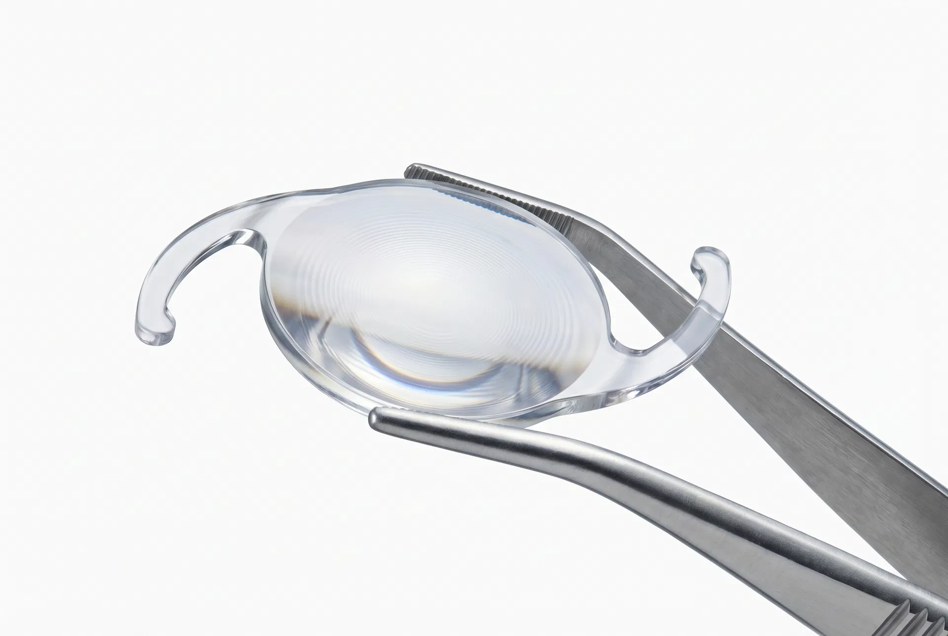
Step 4: IOL Implantation The artificial lens, folded to fit through the small incision, is inserted into the empty lens capsule. It unfolds and positions itself precisely within the capsule.
Step 5: Wound Closure The incisions are self-sealing and typically require no sutures. The eye is protected with a shield.
"The procedure is so refined that most patients are surprised by how comfortable and quick it is. The biggest challenge is usually keeping still for 15 minutes." — Dr. Michael Torres, Cataract Surgeon
Immediately After Surgery
You'll rest in a recovery area for 30–60 minutes while the sedative wears off. Your vision will likely be blurry immediately after surgery — this is normal and expected. You'll be given:
- A protective eye shield to wear (especially while sleeping)
- Antibiotic and anti-inflammatory eye drops
- Detailed post-operative care instructions
- A follow-up appointment for the next day
What to Expect in the First 24 Hours
Most patients experience:
- Mild discomfort, scratchiness, or a foreign body sensation
- Blurry or hazy vision that gradually improves
- Sensitivity to light
- Mild redness or watering
These symptoms are normal and typically resolve within a few days.
Risks and Complications
While cataract surgery is extremely safe, no surgical procedure is without risk. Potential complications include:
| Complication | Frequency | Treatment |
|---|---|---|
| Posterior capsule opacification | 20–40% within 5 years | YAG laser capsulotomy (quick, painless) |
| Refractive error | 5–10% | Glasses, contacts, or enhancement |
| Infection (endophthalmitis) | <0.1% | Urgent antibiotic treatment |
| Retinal detachment | <0.5% | Surgical repair |
| Corneal edema | 1–2% | Usually resolves spontaneously |
The most common "complication" — posterior capsule opacification — is not actually a complication of the original surgery but a natural clouding of the capsule membrane that can occur months to years later. It's easily treated with a quick, painless laser procedure.
The Bottom Line
Cataract surgery is a remarkably safe and effective procedure that restores vision for millions of people each year. Understanding what to expect — from the pre-operative evaluation through the surgical steps — helps reduce anxiety and ensures you're prepared for a smooth recovery.